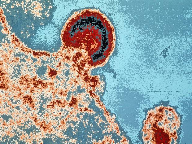
DOES A HIV VIRUS EXIST IN REALITY OR NOT? / By Elena Chobanyan
According to some sources, the basic structure of human immunodeficiency virus type 1 (HIV-1) has been investigated morphologically. But, after all, the HIV-1 core internal structure isn’t finally understood. Few investigations confirmed that a mature HIV-1 particle had two copies of RNA strands in a cone-shaped core. But mIsolated human immunodeficiency virus (HIV) and HIV-infected human lymphocytes in culture have been imaged for the first time by atomic force microscopy (AFM). Purified virus particles spread on glass substrates are roughly spherical, reasonably uniform, though pleomorphic in appearance, and have diameters of about 120 nm. Similar particles are also seen on infected cell surfaces, but morphologies and sizes are considerably more varied, possibly a reflection of the budding process. The surfaces of HIV particles exhibit “tufts” of protein, presumably gp120, which do not physically resemble spikes. The protein tufts, which number about 100 per particle, have average diameters of about 200 Å, but with a large variance. They likely consist of arbitrary associations of small numbers of gp120 monomers on the surface. In examining several hundred virus particles, we found no evidence that the gp120 monomers form threefold symmetric trimers. Although >95% of HIV-infected H9 lymphocytic cells were producing HIV antigens by immunofluorescent assay, most lymphocytes displayed few or no virus on their surfaces, while others were almost covered by a hundred or more viruses, suggesting a dependence on cell cycle or physiology. HIV-infected cells treated with a viral protease inhibitor and their progeny viruses were also imaged by AFM and were indistinguishable from untreated virions. Isolated HIV virions were disrupted by exposure to mild neutral detergents (Tween 20 and CHAPS) at concentrations from 0.25 to 2.0%. Among the products observed were intact virions, the remnants of completely degraded virions, and partially disrupted particles that lacked sectors of surface proteins as well as virions that were split or broken open to reveal their empty interiors. Capsids containing nucleic acid were not seen, suggesting that the capsids were even more fragile than the envelope and were totally degraded and lost. From these images, a good estimate of the thickness of the envelope protein-membrane-matrix protein outer shell of the virion was obtained. Treatment with even low concentrations (<0.1%) of sodium dodecyl sulfate completely destroyed all virions but produced many interesting products, including aggregates of viral proteins with strands of nucleic acid.ore recently, has been shown that the structures of viruses, observed by AFM, are entirely consistent with models derived by X-ray crystallography and cryo-electron microscopy.
 The investigations showed both isolated virions free on glass substrates and virions emerging from infected cell surfaces were imaged by AFM, as were mutant viruses which formed aberrant virions. HIV-infected cells exhibited striking diversity in the number of virus particles present. Only a few virus particles were visible on infected cells. The particles have an average diameter of 127 nm, with a variation of 30 nm, based on measurement of 200 particles. The actual size diversity of the virions is different. Examination of cell membranes from uninfected host cells by AFM reveals a distribution of protein shapes and sizes that are far more diverse than we see on the more or less uniform surfaces of viruses. The tight association that observe between proteins and nucleic acid in the presence of detergents is consistent with the very positively charged nucleocapsid protein binding strongly to RNA. HIV viruses transfer between infected cells through a structure called a virological synapse. When you put all those pictures together, you can watch two cells come together, form a synapse, and then watch the HIV go from one cell to another.
Discussing with Zhaneta Petrosyan, the Head of the Division of Prevention of the Armenian Anti-AIDS Prevention Republican Center, we asked her to find out some details about the HIV virus from another specialist of the center, but she answered that everything was already told.
However, we recently found out what do different people think about the HIV virus.
In Cristian Groza‘s opinion the HIV virus is fake. “People die because of treatment, not because of HIV. HIV doesn’t exist. Does any one have some real images of HIV virus done by a mictoscope..?” – he asked. https://www.youtube.com/user/gman57opPaul M.: „..You know, ebola is more deadly.” Jeff Conforti: “We need to create something that mimics the WBC’s and acts as a decoy attracting the virus to it, instead of the real WBC’s..”
Ivan K. thinks that HIV is not an ilness, which present specialists and the official medicine. HIV isn’t a viral disease, and there is no virus. „The problem is connected to the immune system, which tries to repair damaged cells. If a specialist, doctor, shows me just a HIV virus through a microscope, I will apologize and will admit it. It is obvious that there is an international “network”, which controls every field, branch of people’s life, the medicine as well. We only see the consequences,” – he added.
As we noticed, many people are skeptical about the existence of the virus and are sure that nobody was able to make a real picture of the HIV virus.


I’m really enjoying the theme/design of your site. Do you ever run into any browser compatibility problems? A handful of my blog audience have complained about my site not working correctly in Explorer but looks great in Opera. Do you have any suggestions to help fix this problem?